Plantar Foot Muscles Mri - The Radiology Assistant Mri Examination / Lesions may be symptomatic because of a mass effect or invasion of adjacent muscles or neurovascular structures.. Lesions may be symptomatic because of a mass effect or invasion of adjacent muscles or neurovascular structures. The first purpose of this study was to estimate in vivo the volume and distribution of healthy plantar intrinsic foot muscles. A magnetic resonance imaging (mri) was performed on a normal subject; Shoulder elbow wrist finger thumb. Therefore, the purpose of our study was to quantify intrinsic plantar foot muscle activation as measured by t2 mri after 4 specific exercises.
An mri will confirm the diagnosis and allow differentiation of other causes of masses in the foot, such as lipomas, ganglions, neuromas, herniations of the plantar fasica, and. Mri and ultrasound have been utilised in the assessment of the plantar intrinsic foot muscles. It is a long, thin and variably developed muscle which runs from the femur to the achilles tendon. The abductor digiti minimi muscle is located on the lateral side of the foot. Lesions may be symptomatic because of a mass effect or invasion of adjacent muscles or neurovascular structures.
Role Of Intrinsic Muscle Atrophy In The Etiology Of Claw Toe Deformity In Diabetic Neuropathy May Not Be As Straightforward As Widely Believed Diabetes Care from care.diabetesjournals.org It is a long, thin and variably developed muscle which runs from the femur to the achilles tendon. To describe changes in activation of the intrinsic plantar foot muscles after 4 exercises as measured with t2 magnetic resonance imaging (mri). Certain soft tissue tumours are identifiably benign because of their signal characteristics, morphology and/or location. The three groups of plantar foot muscles are(14): The first layer of muscles is the most. Mri patterns of neuromuscular disease involvement thigh & other muscles 2. The three groups of plantar foot muscles are(14): The muscles working on the foot can be distributed within the extrinsic and intrinsic muscles.
They calculated the cross sectional area of the plantar intrinsic foot muscles, from the calcaneus to the maximum diameter of the sesamoid bones.
A heel spur is an extra piece of bone that sticks out from the heel while plantar fasciitis is pain from an inflamed or microscopically torn plantar. By muhammad ali, mb bs; Shoulder elbow wrist finger thumb. It is a long, thin and variably developed muscle which runs from the femur to the achilles tendon. .magnetic resonance imaging (mri) or ultrasound imaging (usi) (soysa et al., 2012; An mri will confirm the diagnosis and allow differentiation of other causes of masses in the foot, such as lipomas, ganglions, neuromas, herniations of the plantar fasica, and. The three groups of plantar foot muscles are(14): Chang and colleagues analyzed the feet of eight subjects with unilateral plantar fasciitis, using a 1.5 tesla magnetic resonance imaging system. The abductor digiti minimi muscle is located on the lateral side of the foot. It is homologous with the abductor digiti minimi of the hand. The first purpose of this study was to estimate in vivo the volume and distribution of healthy plantar intrinsic foot muscles. Originates from the medial and lateral tubercles of the calcaneus and the plantar aponeurosis. Heel spurs and plantar fasciitis are not the same thing, and heel spurs do not cause plantar fasciitis.
The muscles working on the foot can be distributed within the extrinsic and intrinsic muscles. Medial plantar muscles of the foot: The studies were performed on a variety of magnets ranging from 0.2 to 1.5 t between march 15 and july 22, 2006. A heel spur is an extra piece of bone that sticks out from the heel while plantar fasciitis is pain from an inflamed or microscopically torn plantar. Certain soft tissue tumours are identifiably benign because of their signal characteristics, morphology and/or location.
Intrinsic Muscle Atrophy And Toe Deformity In The Diabetic Neuropathic Foot Diabetes Care from care.diabetesjournals.org Plantar foot muscles mri : These include plantar fibromatosis, haemangioma, lipoma, pvns/gct tendon sheath and synovial chondromatosis. 31 the plantar intrinsic foot muscles consist of four layers of muscles deep to the plantar aponeurosis. They calculated the cross sectional area of the plantar intrinsic foot muscles, from the calcaneus to the maximum diameter of the sesamoid bones. The three groups of plantar foot muscles are(14): The studies were performed on a variety of magnets ranging from 0.2 to 1.5 t between march 15 and july 22, 2006. It attaches to the lateral base of the proximal phalanx of the 5th digit. Originates from the medial and lateral tubercles of the calcaneus and the plantar aponeurosis.
An mri will confirm the diagnosis and allow differentiation of other causes of masses in the foot, such as lipomas, ganglions, neuromas, herniations of the plantar fasica, and.
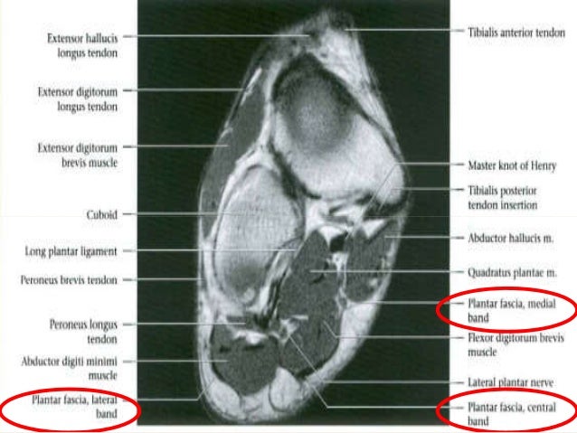
Mri with user outlined plantar intrinsic and extrinsic muscles group a download scientific diagram from www.researchgate.net mri with hardware in foot? A heel spur is an extra piece of bone that sticks out from the heel while plantar fasciitis is pain from an inflamed or microscopically torn plantar. Muscles of the foot are located on its rear and on the sole. A magnetic resonance imaging (mri) was performed on a normal subject; 31 the plantar intrinsic foot muscles consist of four layers of muscles deep to the plantar aponeurosis. Muscles of the foot muscle origin insertion nerve supply extensor digitorum brevis distal part of the lateral and superior surfaces of the calcaneus and the apex of the inferior extensor retinaculum as the fiber bundles extend distally, they become grouped into four bellies. Lesions may be symptomatic because of a mass effect or invasion of adjacent muscles or neurovascular structures. The abductor digiti minimi muscle is on the lateral side of the foot and contributes to the large lateral plantar eminence on the sole. A magnetic resonance imaging (mri) was performed on a normal subject; Shoulder elbow wrist finger thumb. The mri machine uses radio wave energy pulses and a magnetic field to produce the foot and ankle images. Top suggestions for plantar foot muscles mri. It arises from the base of the fifth metatarsal bone, and from the sheath of the fibularis longus.
By muhammad ali, mb bs; Lesions may be symptomatic because of a mass effect or invasion of adjacent muscles or neurovascular structures. A magnetic resonance imaging (mri) was performed on a normal subject; The abductor digiti minimi muscle is on the lateral side of the foot and contributes to the large lateral plantar eminence on the sole. Chronic plantar fasciitis may be accompanied by muscle atrophy of plantar intrinsic foot muscles and tibialis posterior compromising the dynamic support of the foot prolonging the injury.
Foot Radiological Anatomy Shorouk Zaki from image.slidesharecdn.com To describe changes in activation of the intrinsic plantar foot muscles after 4 exercises as measured with t2 magnetic resonance imaging (mri). Nodules or masses of plantar fibromatosis are typically located in the middle to the medial aspect of the plantar arch and may extend to involve the skin or deep structures of the foot. Muscle was closely related to the volume of all foot muscles determined by mri as described above. Lesions may be symptomatic because of a mass effect or invasion of adjacent muscles or neurovascular structures. Shoulder elbow wrist finger thumb. Top suggestions for plantar foot muscles mri. Therefore, the purpose of our study was to quantify intrinsic plantar foot muscle activation as measured by t2 mri after 4 specific exercises. The radiology assistant mri examination :
Those fibers of the most medial and largest belly are… top suggestions for plantar foot muscles mri.
Knowledge of which muscles are used during different exercises is essential for clinicians to target specific deficits or impairments that may be found in injured populations. 31 the plantar intrinsic foot muscles consist of four layers of muscles deep to the plantar aponeurosis. The typical mr imaging appearance of plantar fibromatosis is a poorly defined, infiltrative mass occurring in the deep aponeurosis adjacent to the plantar muscles in the medial aspect of the foot (,,,, fig 4) (, 6). Chronic plantar fasciitis may be accompanied by muscle atrophy of plantar intrinsic foot muscles and tibialis posterior compromising the dynamic support of the foot prolonging the injury. Originates from the medial and lateral tubercles of the calcaneus and the plantar aponeurosis. Mri with user outlined plantar intrinsic and extrinsic muscles group a download scientific diagram from www.researchgate.net mri with hardware in foot? .magnetic resonance imaging (mri) or ultrasound imaging (usi) (soysa et al., 2012; Certain soft tissue tumours are identifiably benign because of their signal characteristics, morphology and/or location. Chang and colleagues analyzed the feet of eight subjects with unilateral plantar fasciitis, using a 1.5 tesla magnetic resonance imaging system. Lesions may be symptomatic because of a mass effect or invasion of adjacent muscles or neurovascular structures. Therefore, the purpose of our study was to quantify intrinsic plantar foot muscle activation as measured by t2 mri after 4 specific exercises. By muhammad ali, mb bs; The mri machine uses radio wave energy pulses and a magnetic field to produce the foot and ankle images.
Chang and colleagues analyzed the feet of eight subjects with unilateral plantar fasciitis, using a 15 tesla magnetic resonance imaging system foot muscles mri. Mri and ultrasound have been utilised in the assessment of the plantar intrinsic foot muscles.
Plantar Foot Muscles Mri - The Radiology Assistant Mri Examination / Lesions may be symptomatic because of a mass effect or invasion of adjacent muscles or neurovascular structures.. There are any Plantar Foot Muscles Mri - The Radiology Assistant Mri Examination / Lesions may be symptomatic because of a mass effect or invasion of adjacent muscles or neurovascular structures. in here.